


Volume 10 • Issue 2
A Growth on the Eyelid:
When to be Suspicious for Skin Cancer

by David R. Jordan
M.D., F.A.C.S., F.R.C.S.(C)
INTRODUCTION
Numerous lumps and bumps can occur on the eyelid or periocular skin as one ages. Some are easily recognizable as benign (xanthalasma, skin tags, inclusion cysts, styes) while others are more difficult to identify and may be benign, precancerous or cancerous.
Skin Cancer is the most frequently occurring neoplasm in the adult population. The head and neck (exposed areas) are common sites for skin cancer to occur. The over-whelming majority of eyelid skin cancers (95%) are Basal Cell Carcinoma followed by Squamous Cell Carcinoma. Eyelid Melanomas are extremely rare as well as other eyelid skin cancer such as Sebaceous Cell Adenocarcinoma, metastatic cancerous lesions and Merkel Cell Carcinoma.
Causes of Skin Cancer
Excessive exposure to sunlight is the single most important factor associated with skin cancers on the face, eyelids and arms. Fair skinned people develop skin cancers far more frequently than dark skinned people. Skin cancer may also be hereditary (ex. Basal Cell Nevus Syndrome). Exposure to chemical carcinogens (ex. arsenic) or ionizing radiation may predispose one to developing skin cancer.
Benign Lumps & Bumps
Benign lid lesions have generally been present for several years. They lie quietly on the skin causing little if any problem. There may occasionally be some mild inflammation (e.g., marginal chalazion) or flaking (some papillomas) but this is the exception rather than the rule. There is rarely if ever, ulceration or bleeding, and growth, for the most part, is very slow, if any occurs at all. Benign lid lesions located along the lid margin do not disrupt the lashes or distort the natural smooth eyelid contour. Some benign lid lesions are pigmented (e.g., an eyelid nevus). When pigmentation is present one should look to see if it is uniform in nature or blotchy and irregular. Benign pigmented lesions usually have a uniform color over their surface.
Malignant Lid Lesions
Eyelid skin cancers occur most often on the lower lid but may also occur on the upper lid, medial or lateral canthal area, eyebrow, or adjacent eyelid skin (periocular region). They usually appear as painless elevated nodules (Figure 1a). There may or may not be some ulceration in the center of the growth (Figure 1b). At times, rather than a nodule, one sees an indentation or erosion of the skin or eyelid margin (Figure 1c). In contrast to the benign lid lesions, there is usually some growth associated with the cancerous or precancerous lesions. Rather than just “sitting there like a bystander” they are growing or changing with time. There may be some flaking, crusting, itching or bleeding. Bleeding is particularly suggestive of a malignancy.
Malignant lesions along the lid margin disrupt the lashes and one may see misdirected lashes or loss of lashes (Figure 1a, 1b, 1c). In addition, the normally smooth eyelid contour becomes irregular in the area of the cancerous growth (Figure 1b,1c).
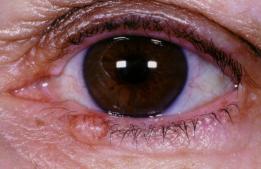
Figure 1a – Basal cell carcinoma on the left lower lid –appearing as a raised, pearly nodule that is disrupting the eyelashes.
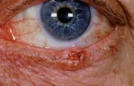
Figure 1b – Basal cell carcinoma on the left lower lid – appearing as a raised lesion with central ulceration. There is eyelash loss and loss of the smooth lid contour.
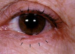
Figure 1c – Basal cell carcinoma on the left lower lid – appears as a poorly defined nodule with areas of lash loss and a distortion of the normal lid contour.
If pigmentation is present, a blotchy, irregular or speckled pattern with light and dark areas is more likely with the malignant lesion than a uniform pigmentation, which is more typical of a benign lesion such as the eyelid nevus.
Although skin cancer is more likely to occur in the aging population, particularly fair skin individuals, it may also occur in younger individuals in the 20’s, 30’s, or 40’s. A lid bump with suspicious features (as described above) in a young (20-40 yrs) individual should be assessed just as it would be in the 60 to 70 year old.
If the patient with a suspicious lid lesion has a past history of having a lid growth removed and it has recurred in the same area, whether it is a few months ago, or 1 to 2 years ago, the suspicion of a cancerous lesion should be raised. In addition, if the patient with a suspicious eyelid bump has a known history of skin cancers occurring in other areas, it increases the likelihood the bump in question is a skin cancer.
SUMMARY
When should you suspect a cancerous eyelid lesion:
Any Bump that:
- is changing in size or appearance
- is itchy and or bleeding
- is associated with some central ulceration or destruction of the skin
- is disrupting, destroying or redirecting the eyelashes
- is disrupting the smooth eyelid contour
- has blotchy, irregular pigmentation
- has regrown after being removed a few months or a few years earlier
- has developed in a patient with other areas of skin a cancer
If you have any questions regarding the topics of this newsletter, or requests for future topics of InSight, please contact Dr. David R. Jordan office by telephone at (613) 563-3800.

