


Volume 4 • Issue 3
Common Eyelid Injuries –Part I

by David R. Jordan
M.D., F.A.C.S., F.R.C.S.(C)
INTRODUCTION
Eyelid injuries may be blunt, penetrating, or avulsive in nature and are often due to flying objects, motor vehicle accidents, tree branches, dog bites cat scratches, etc. The first step in the management of such an injury is to take a careful history in order to appreciate the “mechanism of the injury” and, the “force and velocity” at which the object approached the eye. In all eyelid trauma cases, it is essential to examine the eye in order to rule out damage to the globe prior to treatment of the lid abnormality. A visual acuity examination is essential followed by an assessment of ocular structures and functions (corneal integrity, status of anterior chamber, pupil reaction, ocular motility, etc.)
Blunt injuries
With blunt injuries there is often considerable soft tissue edema and ecchymosis (Figure 1a). This leads to lid swelling which often compromises proper visual assessment. Desmarre retractors or folded paper clips may be used to retract the lids for a more accurate assessment of visual acuity and ocular motility (Figure 1b). It is essential to obtain a visual acuity and record it on the chart. The cornea, anterior chamber, and pupil can be evaluated with a bright, hand-held light. A view of the retina, either with direct or indirect ophthalmoscopy, is often limited but should be attempted at this stage. If the trauma is localized to the soft tissues of the eyelid (ex. hemorrhage and edema), ice packs, can be applied for the first 3 to 5 days (1 hour, 4 times daily) to help reduce swelling. Resolution of the lid edema gradually occurs over 5 to 7 days. Lid ecchymosis usually takes longer. Once the edema starts to settle, a more complete visual assessment can be carried out.
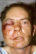
Figure 1a – Soft tissue swelling around eye following a blow. The upper and lower lids are swollen shut.
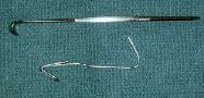
Figure 1b – Desmarre lid retractor above, folded paper clip below, may be used to help open a closed lid.
If ocular motility appears to be abnormal, facial x-rays and tomograms can be used to assess the orbital walls. In cases with ocular motility dysfunction (secondary to a floor or medial wall fracture) we generally wait 12 to 14 days to allow the edema and any possible muscle hemorrhage to settle before considering surgical repair. If continued motility dysfunction exists, entrapment of the muscle or perimuscular tissue (fat and connective tissue) is suspected and a computerized tomography (CT) scan is obtained. Surgery is indicated if there is significant ocular motility impairment, two millimeters or more of enophthalmos, and/or there is a fracture involving more then 50% of the orbital floor or medial wall.
It is not unusual to have a hyphema associated with blunt periocular trauma or a trauma causing a complex mid-facial fracture (zygoma, maxilla, nasal bones). It is important to treat the hyphema first, prior to considering surgical intervention. The risk of a hyphema rebleeding is greatest in the first 72 hours after injury. Any surgery at this time places the eye at increased risk of rebleeding and the potential for serious ocular complications. Thus, if a hyphema is present, we generally wait 7 days to allow its resolution before suggesting repair of extensive mid-facial fractures (which are usually done at day 5 to 7).
A ptotic (dropping) upper eyelid following blunt trauma is best left to resolve spontaneously. Recovery often occurs gradually over a 4 to 6 month period. If the lid remains ptotic at 6 months, ptosis surgery is offered.
Penetrating injuries
For penetrating eyelid injuries (tree branch, knife, etc.), the initial approach remains the same : the eye and various ocular functions must be assessed prior to treatment of the lid defect. Penetrating eyelid injuries may be full or partial thickness, and may include the lid margin and/or canaliculus (Figure 2a). Appropriate x-rays or CT scans are indicated when injury to deeper structures (such as the frontal sinus and orbital rim) are suspected.
For partial-thickness lid lacerations not involving the lid margin (Figure 2b), skin closure alone is sufficient. A 6-0 suture (plain, chromic, silk, nylon) is best for this closure and can be removed in 5 to 7 days (if a non-absorbable suture is used). In children, a 7-0 plain or chromic suture is often the preferred stitch (Figure 2c, d). More extensive partial-thickness lacerations with shredded ends are often seen in the patient who has gone through a car windshield. Gentle cleaning of the wound is recommended in such cases with saline irrigation and removal of foreign bodies (which can also be picked out if embedded). Hydrogen peroxide is excellent to help clean a wound but care should be taken to avoid contact with the cornea. We do not advocate routinely trimming the ends of lacerated lids to obtain “fresh edges” as is often done during the repair of wounds on other parts of the body. The underlying orbicularis muscle is quite vascular and what looks unhealthy initially will often heal normally. Piece-by-piece reapproximation is the preferred technique, using the brow line, lid crease, lash line, and miebomian gland openings as land marks.
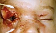
Figure 2a – Penetrating injury of the upper and lower eyelid following a motor vehicle accident.
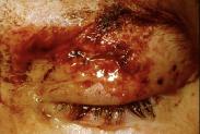
Figure 2b – Partial thickness lid laceration.
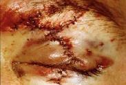
Figure 2c – Meticulous closure with a fine absorbable suture.
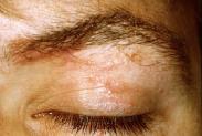
Figure 2d – Results after sutures have absorbed.
Avulsive injuries
Avulsive eyelid injuries involve tissue loss and planning of appropriate lid reconstruction. Penetrating injuries due to sharp objects usually leave all the tissue present, whereas dog bite injuries often have areas of missing tissue (Figure 3a). The lacerated lid can be dealt with in a similar fashion to the eyelid undergoing reconstruction following tumor resection. The tarsus and conjunctiva form the “posterior lamella”, while the skin and orbicularis form the “anterior lamella”. Both anterior and posterior lamella require reconstruction. One should try and replace the missing tissue with tissue of the same consistency. It is important to remember during lid reconstruction that a graft on top of a graft will not survive. Thus, one lamella must always have an intact blood supply while the other lamella can be a free graft without an intact blood supply.
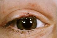
Figure 3a – Central portion of lid margin is missing (absent area of lashes) following a dog bite injury.
Anterior lamellar reconstructive techniques may involve mobilization of surrounding tissue, myocutaneous flaps, pedicle or rotational flaps, and skin grafts. Posterior lamellar reconstructive procedures include free tarsal grafts, sliding tarsoconjunctival flaps, use of tarsal substitutes (e.g. nasal or ear cartilage, hard palate mucosa, banked sclera, and composite grafts from the opposite eyelid. Various eyelid sharing procedures, such as the Cutler-Beard and Hughes techniques, are other well-described reconstruction techniques.
Avulsive injuries with tissue loss can be difficult to repair and a sound knowledge of the lid anatomy as well as various reconstructive techniques is required to achieve a successful result.
If you have any questions regarding the topics of this newsletter, or requests for future topics of InSight, please contact Dr. David R. Jordan office by telephone at (613) 563-3800.

