Volume 3 • Issue 4
Common Eyelid Lumps and Bumps

by David R. Jordan
M.D., F.A.C.S., F.R.C.S.(C)
INTRODUCTION
Numerous benign lumps and bumps can occur on the eyelid and periocular region with increasing age. Generally, benign lid lesions have been present for at least 6 months, are lying quietly on the skin causing little, if any, problem. Benign lid lesions usually do not disrupt the lashes or distort the smooth lid contour. A history of increasing growth, bleeding, ulceration, change in color, recurrence of the lesion after previous removal are historical features of importance that may suggest a malignant growth. A lid growth causing lash loss or a disruption of the normal lash line and/or is distorting the natural smooth lid contour should raise ones suspicions of malignancy.
Hordeola (Styes) and Chalazions
An external hordeoleum (stye) results from an acute purulent inflammation of the superficial sweat glands, sebaceous glands or hair follicle of the eyelids, while an internal hordeolum occurs in the miebomian glands within the tarsal plates of the lids.
A chalazion is a chronic inflammation of a miebomian gland (deep type) or Zeiss sebaceous gland (superficial type) resulting in a clinically firm, painless nodule of the eyelid.

Figure 1 – Chalazion
Treatment of the acute phase may involve hot compresses, antibiotics (drops, ointment, oral) while treatment of the chronic phase usually involves incision and drainage.
Xanthelasma
Xanthalasma are superficial yellowish deposits commonly seen in the medial aspect of the upper and lower lids. It is unclear what triggers their onset. They represent collections of lipid material in the superficial dermis. Blood cholesterol and lipid levels should be checked as 30%will have an elevated lipid level that require treatment. Xanthalasma can be treated by chemicals, surgical excision or laser. Approximately 30% of them will recur.
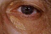
Figure 2 – Xanthelasma
Epidermal Inclusion Cysts
These are small white-yellow cystic lesions occurring on the lid skin, conjunctiva, face or neck and are very common. They may develop spontaneously or arise following trauma or after surgery along an incision line. They originate from pilosebaceous follicles or invagination of surface epidermis. Some of these cysts occurring amongst the lashes will be difficult to distinguish from a blocked Zeiss gland (sebaceous gland). Treatment involves excision with a small scissors or simply unroofing them with a 20 gauge needle. If the cyst wall is not removed, recurrence may occur.

Figure 3 – Inclusion Cysts
Sebaceous Cysts
Sebaceous cysts may also occur in the aging patient around the eyelid area and clinically resemble epidermal inclusion cysts. They are generally found in locations with many hair follicles, particularly the brow area and medial canthus. These cysts may occur secondary to obstruction of the Zeiss gland, miebomian gland or sebaceous glands associated with hair follicles of the lid skin or brow area. Unlike an epidermal inclusion cyst (filled with keratin material), a sebaceous cyst contains epithelial cells, keratin, fats and cholesterol crystals. Treatment involves surgical excision.

Figure 4 – Sebaceous Cyst
Milia
Milia are multiple well-delineated, round, yellow-white cystic lesions ranging from 1 to 3 mm in diameter, found on the face, lids, cheeks and nose. They may occur spontaneously or arise following trauma. They are felt to be retention follicular cysts caused by blockage of the fine pilosebaceous units (hair follicle). Surgical excision is the treatment of choice.
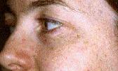
Figure 5 – Milia
Sudoriferous Cysts, (Retention Cysts, Hidrocystomas)
The lid skin has numerous sweat glands (eccrine glands) and modified sweat glands (apocrine glands) such as the glands of Moll. Blockage of these glands leads to the formation of a translucent (water blister-like) lesion on the lid skin or lid margin amongst the lashes. They may occur as a single lesion or as several. Treatment involves total excision other-wise they will recur. Simply stabbing them with a needle is ineffective in allowing their resolution.

Figure 6 – Sudoriferous Cyst (retention cyst, hidrocystoma)
Papillomas
Papillomas are the most common benign lesion of the eyelid. The exact etiology is unclear. These growths represent a benign hyperplasia of the surface epithelium and may be sessile or pedunculated. They occur in middle aged and elderly individuals and may be solitary or multiple, occurring any where on the eyelid. Papillomas differ from the infective warts which consist of inflammatory hyper-trophy of surface epithelium with viral inclusions. Treatment is by surgical excision or laser ablation.
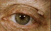
Figure 7 – Papillomas
Seborrheic Keratosis
Seborrheic keratosis are a common benign lesions on the lids of aging individuals. They are well circumscribed, waxy, friable and appear stuck on to the skin. The lesion is very superficial and may be pigmented. Microscopically, they show hyperkeratosis and cystic areas filled with keratin. Their etiology is unclear. Treatment involves surgical excision or laser ablation.
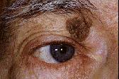
Figure 8 – Seborrheic Keratosis
Nevi
Nevi are common in the eyelid area and may be pigmented or non-pigmented. The nevus consists of a collection of benign appearing dermal melanocysts. Generally, they have been present for years with no change. If the duration of any pigmented lesion is unknown, acquired with age (especially after 40) or there is a change in size or color, a biopsy is recommended.

Figure 9 – Nevus
Pyogenic Granuloma
Pyogenic granuloma is a common vascular lesion of the eyelid that occurs after trauma or surgery. Clinically, it is a fast-growing pinkish mass with occasional superficial ulceration of the epithelium. They are usually pedunculated and often cause discharge when located on the conjunctival surface. Microscopically, they are essentially excess healing tissue (granulation tissue) that occur at sites of irritation (trauma or surgical incisions). They have a rich supply of capillaries that spread from the base of the lesion to the surface.

Figure 10 – Pyogenic Granuloma following ruptured chalazion
In summary, there are numerous benign lumps and bumps that may occur in the aging population, several of which have a characteristic presentation and appearance. Benign lesions generally do not disrupt or distort the lash line or lid contour. They tend to lie quietly on the skin or near its surface with little growth or color changes. Malignant lesions tend to grow, distort, and disrupt the lash line and lid contour and may change in appearance, color and surface texture. They tend to invade surrounding tissues and disrupt their natural appearance. It is important to bear in mind that malignancies can occasionally mimic the benign lesions. Early diagnosis requires an accurately taken history, a high index of suspicion and a biopsy when uncertain.
If you have any questions regarding the topics of this newsletter, or requests for future topics of InSight, please contact Dr. David R. Jordan office by telephone at (613) 563-3800.


