


Volume 5 • Issue 2
Disorders of the Eyelashes

by David R. Jordan
M.D., F.A.C.S., F.R.C.S.(C)
INTRODUCTION
Inturned eyelashes (trichiasis) are a common source of recurrent ocular irritation for some patients. The inturned lash (or lashes) in contact with the conjunctiva and/or cornea may lead to a foreign body sensation, localized conjunctival injection, pain and photophobia. Management of the inturned eyelashes can be challenging for the physician, as well as frustrating for the patient, particularly if recurrent visits are required to eliminate them.
What causes trichiasis?
Trichiasis (Figure 1) is the term used for misdirection or aberrant placement of eyelashes along the eyelid margin resulting in lash growth toward the cornea. Trichiasis is an acquired condition that may be caused by the following inflammatory or traumatic processes involving the eyelids:
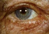
Figure 1– Inturned eye lashes on lower lid.
Chronic blepharitis with meibomianitis – chronic inflammatory changes within the tarsal plate and posterior eyelid margin may cause destruction and misdirection of lash follicles, resulting in
chronic trichiasis.
Lid lacerations and thermal burns to the lid margin – may cause redirection of the lash roots with resultant trichiasis.
Previous surgery on eyelids – For example, lid adhesions (tarsorrhaphys) done to prevent exposure in some patients with seventh nerve palsys may cause misdirection of lashes. Similarly, in many reconstructive eyelid procedures, the new eyelid margin may contain fine skin hairs (lanugo-type) that rub on the cornea.
Mucocutaneous diseases – Stevens-Johnson Syndrome and Ocular Cicatricial Pemphigoid result not only in the destruction of the eyelid margins and trichiasis but also in the formation of new lashes from the meibomian gland orifices (a condition referred to as distichiasis (Figure2).
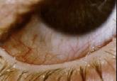
Figure 2– An accessory row of eyelashes is seen, sprouting from the meibomian gland openings (distichiasis).
Other cicatricial conjunctival diseases –Herpes Simplex conjunctivitis and Herpes Zoster may cause a cicatrizing conjunctivitis with destruction of the lid margin and lash follicles. Trachoma may also cause a chronic tarsitis with cicatrizing conjunctivitis in the upper or lower eyelid and resultant trichiasis (as well as a cicatricial entropion).
Irradiation and chemical burns–Therapeutic irradiation for eyelid cancers or alkali burns may lead to a disruption of the normal eyelid margin anatomy and resultant misdirection of eyelashes. Both of these processes may also lead to metaplasia of squamous epithelium of the mucocutaneous margin of the eyelid with resultant keratinization, a source of ocular irritation. In addition, destruction of the goblet cells, accessory lacrimal glands, and lacrimal gland will disrupt the normal tear f low, com-pounding the above problems.
What is the differential diagnosis?
Trichiasis must be differentiated from the following:
Eyelid entropion – True entropion (e.g. involutional type seen in the aging population) is characterized by a normal eyelid margin architecture: the eyelid inverts as a result of eyelid laxity, allowing the eyelashes to rub on the cornea. Several of the entities mentioned above (Ocular Pemphigoid, Stevens-Johnson Syndrome) may cause a cicatrization of the conjunctiva as well as the lid margin and create a cicatricial entropion with trichiasis (i.e. the eyelid is inverted due to a cicatricial process). In addition, eyelashes may be misdirected not only due to the lid position, but also due to the inflammatory process involving the actual lash follicles. Therefore, sometimes there may be two problems present (entropion and trichiasis) both of which may require treatment.
Epiblepharon – Epiblepharon is a congenital condition commonly seen in the lower Asian eyelid. A fold of skin and muscle roll upwards and press the lashes toward the cornea (Figure 3). This does not represent true trichiasis.
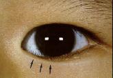
Figure 3– A roll of skin (arrows) is pushing lashes vertically and toward cornea.
Distichiasis – is an abnormality in which an aberrant second row of lashes, (usually from the meibomian gland orifices) grows behind the normal lash line (Figure 2). It may be congenital or acquired. Any process causing chronic inflammation of the lid margin and meibomian glands may transform the meibomian glands into pilosebaceous units capable of producing hair (e.g. chronic blepharitis).
Combined eyelid margin process – Several of the eyelid processes mentioned (Stevens-Johnson Syndrome, Ocular Pemphigoid, irradiation, chemical burns) not only may cause entropion and trichiasis, but in addition may lead to squamous metaplasia and keratinization of the non-keratinizing squamous epithelium of the eyelid margin (Figure 4). Keratinized tissue is very irritating to the eye. Therefore, several factors may contribute to the ocular irritation, and as a result, several types of treatment could be required.
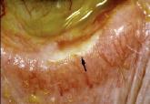
Figure 4– An area of Keratinization (arrow) is seen along the conjunctival side of the lid margin.
Marginal entropion – Is a subtle form of entropion that is seen only at the lid margin. Usually there is chronic inflammation at the eyelid margin with a mild cicatricial process that is starting to roll the lid margin inward. The eyelashes appear more vertical with some truly trichiatic lashes. The clinical clue is the meibomian gland orifices. Normally they should be vertical and not covered by conjunctival epithelium. If the openings are rolled inward and conjunctiva is growing over the opening, then marginal entropion is present in addition to trichiasis. It is important to distinguish this condition when considering treatment.
Diagnostic tips
During the initial patient interview, there may be a history of previous surgery, chemical burns, trauma, etc. that may offer an explanation for the trichiatic lashes.
On examination, look at the lashes carefully. Are there one or two lashes turned in, or a clump of 5 or 6? Are there signs of blepharitis and meibomianitis? Are the meibomian glands rolled in and covered with conjunctiva? Are any signs of conjunctival scarring seen? Is the scarring localized to the area of misdirected lashes or all along the conjunctival surface? Is there keratinization along the lid margin? Is there any shortening of the fornices? Do any lashes grow out of the meibomian glands?
Answering these questions should help decide what type of disease process is responsible for the inturned lashes. Establishing the correct etiology of misdirected lashes is important because it influences therapy and prognosis. For example pemphigoid presents a special problem as the treatment for the inturned lashes may affect the actual disease process.
Treatment
The goals of treatment are 1) preservation of vision, 2) comfort, and 3) acceptable appearance of the lid.
Eyelash disorders can be managed in one of three ways; 1) epilation, 2) destruction of offending lashes and their follicles, or 3) surgical alteration of the eyelid. Selection of the appropriate management approach depends on etiology, ex tent of the abnormality, location of the lashes and the needs of the individual patient.
Epilation – Some patients find simple epilation a reasonable treatment. If the same lashes recur repeatedly – a more permanent solution should be sought.
Destruction of the lash follicle – A variety of procedures are available to destroy the lash follicles including: electrolysis, Argon laser ablation, and cryotherapy.
Electrolysis – local anesthesia is required. The tip of a fine electrode is inserted into the follicle opening. A very small electrical current is applied until bubbling is noted at the follicular orifice. This is best used for isolated (1-4) lashes. Complications are few with recurrence being the most common complication.
Argon laser – lashes are individually treated with the Argon laser under local anesthesia (Figure 5). Laser settings of 1.5 watts, 100 micron spot size for 0.5 seconds to a depth of 2 mm using the yellow/green or blue setting works well for isolated (1-8) lashes. Complications are few and include recurrence and dimpling at the lash site.
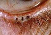
Figure 5– Inturned eyelashes have been lasered away (black spots).
Cryotherapy – Cryo kills cells by causing intracellular ice crystal formation (Figure 6). Lash follicles appear to be more sensitive to the effects of freezing then surrounding lid tissue. The process is done under local anesthesia and is often used for clumps of inturned lashes (5-7) on the lid. Complications include an inflammatory response that may aggravate the process causing trichiasis (e.g. pemphigoid), loss of pigmentation, misdirected lashes adjacent to the cryo site, and recurrence.
Surgery
Surgical procedures involve either repositioning or removing the lash line.
Repositioning procedures – may involve splitting the eyelid and recessing the misdirected lashes. A mucosal graft or hard palate graft may be used during the procedure. Another repositioning procedure involves incising the tarsal plate along its entire length (parallel to the lid margin) and then closing the lid defect in such a way that the eyelids are rotated outward (tarsotomy).
Removal procedures – a full thickness resection of the lid where the inturned eyelashes are located may be useful to remove offending lashes in some patients.
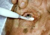
Figure 6-Cryo is being applied to area of inturned eyelashes.
In summary, inturned eyelashes are common and have a variety of causes. Diagnosis of the actual cause is essential. Each case is considered on an individual basis and appropriate treatment is selected for the specific problem at hand. Treatment at times can be frustrating since it may generate its own set of problems including recurrence, lid scarring, loss of lid tissue, deformity of lid tissue, and exacerbation of ocular surface problems. Chronic, progressive disease associated with recurrent lash disorders may require continuing management and may resist permanent cure.
If you have any questions regarding the topics of this newsletter, or requests for future topics of InSight, please contact Dr. David R. Jordan office by telephone at (613) 563-3800.

